fellow Ota Memorial Sleep Center kawasakicity, Kanagawa, Japan
Disclosure(s):
SHINTARO CHIBA: No relevant relationships to disclose.
Kazumi Okuno, MD: No relevant relationships to disclose.
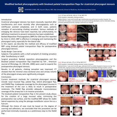
Introduction: Cicatricial pharyngeal stenosis has been classically reported after tonsillectomy and more recently after pharyngoplasty such as LAUP(Laser assisted uvloplasty), and in both cases, patients complain of excruciating choking sensation. Various methods of enlarging the stenosis have been reported, but unfortunately, no definitive treatment to prevent restenosis has been established. Barbed Reposition Pharyngoplasty (BRP) for OSA was first reported by Vicini in 2015; BRP is effective in enlarging and maintaining the pharyngeal cavity, especially for the lateral side. In this report, we describe the method and efficacy of modified BRP using bivalved palatal transposition flaps for postoperative pharyngeal stenosis.
Methods: Study design: Case series Subjects: Five patients with a chief complaint of choking sensation after Tonsillectomy or LAUP. Surgical procedure: Barbed reposition pharyngoplasty and the Bivalved palatal transposition flap (reported by Toh , American Journal of Rhinology 14, 199-204, 2000) were used to expand the postoperative pharyngeal stenosis.
Results:
Results: In all patients, choking sensation was improved. CT showed that the minimal cross-sectional area and airway volume of the pharyngeal airway were significantly enlarged.
Conclusions: Current treatment methods for cicatricial pharyngeal stenosis include nasal mucosal flap, palatal flap, lateral pharyngeal flap, and FAMM flap. Inadequate coverage by the pharyngeal mucosa in the treatment of the scar is likely to result in postoperative restenosis. The FAAM flap provides adequate mucocutaneous coverage of the airway but is an invasive procedure. The bivalved palatal transposition flap in the present study allows for the creation of a large mucosal valve, minimizing the postoperative raw surface, and the modified BRP allows for stable lateral expansion by using the pterygo-mandibular suture line as a fulcrum. Although the choice of case must be based on the degree of scarring and adhesions, we concluded that this procedure can be performed minimally invasively as a preliminary step to the FAAM flap.